Scientists have identified a new way to detect signs of motor neuron disease (MND) in brain tissue, raising hopes for early diagnosis and intervention before onset of symptoms.
Researchers used a molecule known as an aptamer, an approach which has already been applied in cancer diagnostics, to detect MND in brain tissue samples.
The test would need further development before it could be used in clinics. However, scientists predict the new aptamer, TDP-43, could trigger a step-change in MND research, supplementing or replacing some traditional antibody approaches for detection.
“With better ability to detect disease we might be able to diagnose people with MND earlier, when therapeutic drugs might be much more effective,” said Dr. Holly Spence, a co-author of the study and University of Aberdeen research fellow.
The findings were reported in Acta Neuropathologica by researchers from Aberdeen University, the University of Edinburgh, and international partners.
In motor neuron disease, also known as amyotrophic lateral sclerosis (ALS), the accumulation of certain proteins in the brain clump together, causing the cells to gradually stop working. As the disease progresses, it impairs movement, thinking, and breathing, all of which worsen over time.
TDP-43 is an RNA-binding protein, the accumulation of which is a hallmark of ALS and related MNDs. In the latest study, researchers developed a novel RNA aptamer, TDP-43APT, to detect TDP-43 aggregation by binding to protein clumps in the brains of people with MND.
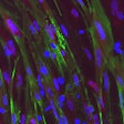
It was able to identify damaged proteins in brain tissue samples before the cells malfunctioned – the stage at which MND symptoms appear and current tests identify the disease. It detected accumulating proteins at much lower levels and with higher accuracy than existing approaches and revealed that toxic protein clumps are building up well before people show symptoms.
“We demonstrate that TDP-43APT identifies pathological TDP-43, detecting aggregation events that cannot be detected by classical antibody stains,” the researchers wrote.
“The improved sensitivity and specificity of TDP-43APT in detecting TDP-43 pathology, if adapted as a PET tracer or liquid biomarker, could aid in the identification of individuals for targeted early interventions,” they note.
Dr. Mathew Horrocks, a co-author and senior lecturer in biophysics, at the University of Edinburgh, said it was the first time the TDP-43 aptamer had been used in human tissue.
“We’re excited by its ability to detect a pathological form of the protein that has so far been difficult to characterize,” he said.
The researchers say RNA aptamers can be made quickly and easily in a test tube without relying on using animals to generate antibodies.
“That means we can really scale this up,” Dr. Jenna Gregory, principal investigator, from the University of Aberdeen, told BBC Radio.
“We really need to be doing more for people affected by MND. In my mind the earlier we intervene the more successful we are likely to be with therapies, that’s exactly what we’re doing with this tool,” she said.
A therapeutic, Tofersen, has recently been licensed in the U.S. and Europe for a genetic cause of MND; scientists say it is increasingly important to develop biomarkers to detect disease in tissue.
Dr. Brian Dickie, director of research at the Motor Neurone Disease Association, said, “It often takes a year from the first onset of symptoms to receiving a diagnosis of MND. This innovative research into the early cellular changes occurring in MND offers exciting potential for the development of new tests to help reduce diagnostic delay. As treatment does not begin until the disease is diagnosed, earlier intervention will hopefully also mean that treatments are more effective."


