Researchers have developed soft, miniaturized wearable devices to continuously capture the sounds of breathing and other bodily functions.
The sounds of air moving through the lungs, the heart beating, and food passing along the digestive tract can provide insights into the health of an individual. However, traditional stethoscopes only capture the sound in a small area of the body at a particular time. Devices that collect sound data continuously from a larger area of the body may yield additional insights to aid in improving the diagnosis and monitoring of health conditions.
That idea underpins a paper published Thursday in Nature Medicine. In the paper, Northwestern University researchers describe the development and assessment of digital microphones and accelerometers that affix to the skin to form a network of sensors that continually capture sound data.
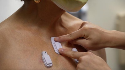
Each device features a flash memory drive, battery, electronic components, Bluetooth capabilities, and two microphones encapsulated in silicone. One microphone faces inward to capture sounds from the body. The other microphone faces outward to collect ambient sounds, providing data that an algorithm can use to determine whether a sound came from the patient or an external source.
“Lungs don't produce enough sound for a normal person to hear. They just aren’t loud enough, and hospitals can be noisy places. When there are people talking nearby or machines beeping, it can be incredibly difficult. An important aspect of our technology is that it can correct for those ambient sounds,” Dr. Ankit Bharat, a thoracic surgeon at Northwestern Medicine, said in a statement.
Bharat led one of two clinical assessments of the technology. In the study led by Bharat, 35 adults with chronic lung diseases and 20 healthy controls wore the sensors. Placing the sensors, which are 40 mL long, 20 mL wide, and 8 mm thick, at multiple locations across the chest enabled the researchers to track each breath across different regions of the lungs.
The other study placed sensors on 15 premature babies in a neonatal intensive care unit with respiratory and intestinal motility disorders. Applied to four parts of the abdomen, the sensors captured results that aligned with measurements of adult intestinal motility using wire-based systems.
“When placed on the abdomen, the automatic detection of reduced bowel sounds could alert the clinician of an impending, sometimes life-threatening, gastrointestinal complication. Improved bowel sounds could indicate signs of bowel recovery, especially after a gastrointestinal surgery,” Dr. Wissam Shalish, a neonatologist at the Montreal Children’s Hospital, said.
The researchers also put the sensors near the base of the throat to detect the presence of airflow and chest movements in babies. The sensors enabled the team to estimate the level of airflow obstruction and thereby identify and classify subtypes of apnea, a leading cause of prolonged hospitalization in premature babies.


