Researchers are developing 3D spatial maps that provide intricate information about the biological features driving the growth of tumors.
One of the most recent examples is part of a comprehensive atlas of colorectal cancer published early this month in Cell and described on Tuesday by Lawrence Tabak, acting director of the National Institutes of Health (NIH), in the NIH Director’s Blog.
“Working this ‘molecular information’ into the pathology report would bring greater diagnostic precision, drilling down to the actual biology driving the growth of the tumor," Tabak wrote. "It also would help doctors to match the right treatments to a patient’s tumor and not waste time on drugs that will be ineffective."
The colorectal cancer atlas described in Cell was developed by an NIH-supported team led by Sandro Santagata, Brigham and Women’s Hospital, Boston, and Peter Sorger, Harvard Medical School, Cambridge, MA, in collaboration with investigators at Vanderbilt University, Nashville, TN.
They had previously described a high-definition map of melanoma, which was published last June in Cancer Discovery.
Both atlases are part of the Human Tumor Atlas Network supported by the NIH’s National Cancer Institute.
“What’s so interesting with the colorectal atlas is the team combined traditional pathology with a sophisticated technique for imaging single cells, enabling them to capture their fine molecular details in an unprecedented way,” Tabak added.
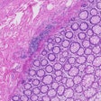
The team leveraged cyclic immunofluorescence, which uses many rounds of highly detailed molecular imaging for each tissue sample to generate molecular data, cell by cell. The researchers captured this information for almost 100 million cancer cells isolated from tumor samples representing 93 individuals diagnosed with colorectal cancer.


