A hospital in São Paulo used a portable ultrasound machine to perform autopsies on 10 patients with fatal cases of COVID-19. The authors described the modality's benefits for studying the effects of the disease caused by the novel coronavirus in a paper published on May 22 in Histopathology.
Using a technique called ultrasound-based minimally invasive autopsies (MIA-US), an ultrasound examiner and technologist took tissue samples from the most affected parts of each patient's organs. The findings confirmed that COVID-19 affects multiple organs and tissues, including the kidneys, spleen, lymph nodes, brain, testicles, and skin.
"MIA-US provides fast, precise responses to investigate the pathogenesis of new infectious agents and, thus, represents a valuable alternative to conventional autopsies in situations of high contagiousness," wrote the team of Brazilian authors, led by Dr. Amaro Nunes Duarte-Neto and Dr. Renata Aparecida de Almeida Monteiro from the Brazilian Image Autopsy Study Group at the University of São Paulo medical school.
Ultrasound-guided autopsies had previously been used on patients during a 2018 yellow fever epidemic in São Paulo. The authors used the same techniques on the first 10 patients who died from COVID-19 at their hospital in March of this year. The patients all had underlying conditions, including hypertension, diabetes, and advanced age.
The hospital took a number of precautions to protect the ultrasound examiner and technologist who conducted the autopsies, including covering the deceased in multiple plastic bags. The ultrasound team also wore head-to-toe personal protective equipment, including two aprons, rubber boots, plastic sleeves, three glove layers, a rubber cap, an N95 mask, a surgical mask, and eye protection.
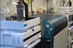
During the autopsies, the examiner and technologist used a portable ultrasound machine with multifrequency broadband transducers and standard image quality. They scanned patients' organs to identify the most-affected locations, then cut 10-cm openings at the appropriate locations to take ultrasound-guided tissue samples from the lung, liver, kidneys, spleen, and heart. They also took unguided samples from the quadriceps, skin, and brain.
The samples revealed significant lung findings, including exudative and/or proliferative diffuse alveolar damage. The authors also found severe alveolar epithelial changes, which they described as more intense and prevalent than findings for other respiratory viruses.
Many but not all findings outside of the lungs could be attributed to the comorbidities of the patients or to septic shock, the authors noted. For instance, eight patients had fibrinous thrombi in alveolar arterioles and a high density of alveolar megakaryocytes, which the authors believed could be evidence of a hypercoagulative state in severely ill patients. The researchers also found cases with superficial perivascular dermatitis, myositis, orchitis, and myocarditis.
The hospital's staff has since taken into account the autopsy findings when addressing other critically ill patients with COVID-19, the authors noted. In addition, no one on the ultrasound team tested positive for COVID-19 in the 15 days following the autopsies.
It is also important to note that diagnostic testing only identified COVID-19 in nine of the 10 patients in the study. However, the authors strongly believed that the tissue samples and clinical information from the tenth patient matched the others, so they provided a COVID-19 diagnosis in the absence of a positive test result.
"Other causes of respiratory failure may be associated with or simulate COVID-19, and a [noninvasive] diagnostic approach, such as naso/oropharyngeal [test], can generate a large number of false negatives," the authors wrote. "This issue is even more critical in cases of death, where the actual mortality rate can be underestimated."