Researchers have designed a high-speed 3D microscope that is able to see real-time cellular detail in living tissues, allowing for the label-free imaging of diagnostically relevant tissue, according to research published on March 28 in Nature Biomedical Engineering.
The system, called MediSCAPE (Swept Confocally Aligned Planar Excitation microscopy), is capable of capturing images of tissue structures that can guide surgeons to navigate tumors and their boundaries without needing to remove tissues and wait for pathology results. If proven in the field, the technology could eventually replace conventional biopsies and histology with real-time imaging within the living body.
"The way that biopsy samples are processed hasn't changed in 100 years," said Elizabeth Hillman, PhD, professor of biomedical engineering and radiology at Columbia University and senior author of the study, in a statement. "They are cut out, fixed, embedded, sliced, stained with dyes, positioned on a glass slide, and viewed by a pathologist using a simple microscope. This is why it can take days to hear news back about your diagnosis after a biopsy."
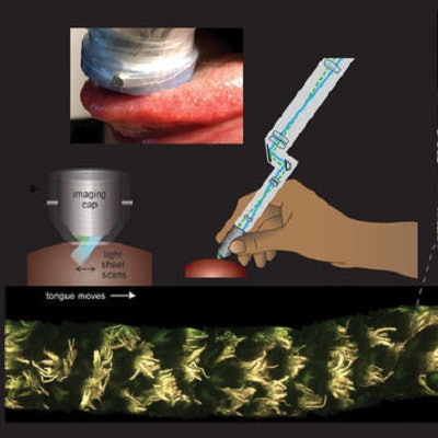
 MediSCAPE imaging of the living human tongue. Subject slowly licked the MediSCAPE imaging head while high-speed 3D images were acquired. Volumes were then stitched to form long 3D strip. Contrast shows red and green autofluorescence with 488 nm excitation. Inset left shows MediSCAPE imaging geometry in which an oblique light sheet is used to illuminate the sample, and scans from side to side to rapidly capture 3D images. Inset right shows concept of handheld MediSCAPE system for intrasurgical use.
Like scanning with a flashlight
Over the past decade, Hillman and her students had been developing new kinds of microscopes that can capture fast 3D images of living samples like tiny worms, fish, and flies.
"One of the first tissues we looked at was fresh mouse kidney, and we were stunned to see gorgeous structures that looked a lot like what you get with standard histology," said Kripa Patel, PhD, a recent graduate from the Hillman lab and lead author of the study.
The microscope, which they named SCAPE, was able to provide brightly illuminated color views without adding any dyes to the mouse tissue.
"Everything we saw was natural fluorescence in the tissue that is usually too weak to see," Patel said. "Our microscope is so efficient that we could see these weak signals well, even though we were also imaging whole 3D volumes at speeds fast enough to rove around in real time, scanning different areas of the tissue as if we were holding a flashlight."
From mouse kidneys to human kidneys
For the next experiment, this time on human tissues, the team obtained fresh samples from human kidneys. As in the mouse experiments, the microscope was able to detect telltale signs of kidney disease that matched well to conventional histology images.
The team's next goal was to test the technology on live human organs in the body. This required reducing the footprint of the large SCAPE microscopes in Hillman's lab to something that would fit into an operating room and could be used by a surgeon. The smaller version of the system was dubbed MediSCAPE.
When MediSCAPE was tried on a live human kidney in a live body, it allowed the researchers to visualize blood flow through the tissues and see the cellular-level effects of ischemia and reperfusion, a pathological condition characterized by an initial restriction of blood supply followed by its subsequent restoration and reoxygenation.
"Understanding whether tissues are staying healthy and getting good blood supply during surgical procedures is really important," says Hillman. "We also realized that if we don't have to remove (and kill) tissues to look at them, we can find many more uses for MediSCAPE, even to answer simple questions such as 'what tissue is this?' or to navigate around precious nerves."
No more biopsies?
A biopsy, which requires cutting out small pieces of tissue for examination under a microscope, presents doctors with a number of challenges, particularly when the tissue is in a sensitive area of the body such as the brain, spinal cord, nerves, eye, and areas of the face.
"Because we can image the living tissue without cutting it out, we hope that MediSCAPE will make those decisions a thing of the past," Hillman said.
Hillman added that the technology should be particularly useful for robotic and laparoscopic surgeries where surgeons are more limited in their ability to identify and interact with tissues directly.
The team is currently organizing a large clinical trial with the goal of commercialization and U.S. Food and Drug Administration approval.