Business Insights
Diagnostic Technologies
Diseases
Point-of-Care Testing
Research & Development
Sign In
Clinical Chemistry
Emerging Technology
Immunoassays
Molecular Diagnostics
Pathology & AI
Microscopy & Imaging: Page 2
U.K. pathology group expands use of Sectra software
The NPIC said this expansion will help laboratories in different parts of the U.K. with these efforts. It is a result of a five-year contract signed in October 2021 and is part of a multimillion-pound National Health Service initiative.
March 3, 2022
Spinal fluid tests may aid PET scans of Alzheimer's disease
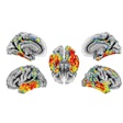
A Swiss group led by senior author Michael Schöll, PhD, of the University Gothenburg investigated whether fluid and blood biomarkers that indicate Alzheimer's disease could predict the accumulation of neurofibrillary tau tangles seen on PET scans in patients diagnosed with the disease. They found the lab tests performed well enough to help select patients for PET imaging studies evaluating new treatments.Associations of baseline beta-amyloid PET and baseline phosphorylated tau-181 (p-tau181) with F-18 flortaucipir (FTP) Standardized uptake value ratio (SUVR) annualized change for (A) cognitively unimpaired and (B) cognitively impaired individuals. Image courtesy of Alzheimer's & Dementia: The Journal of the Alzheimer's Association.Associations of baseline beta-amyloid PET and baseline phosphorylated tau-181 (p-tau181) with F-18 flortaucipir (FTP) Standardized uptake value ratio (SUVR) annualized change for (A) cognitively unimpaired and (B) cognitively impaired individuals. Brain surface renderings depict Pearson correlation coefficients adjusted for age and sex (r) representing the strength of the association between the different biomarkers and F-18 flortaucipir SUVR change in each brain region. The right upper panel represents age- and sex-adjusted Pearson correlation coefficients (r), along with p-values, for the association between the biomarkers and F-18 flortaucipir SUVR change in the Alzheimer's disease meta-region of interest (ROI). The root mean squared error (RMSE) for the prediction of F-18 flortaucipir SUVR change by a linear model with the biomarker as predictor and age and sex as covariates. RMSE was estimated using leave-one-out cross-validation. Voxel-wise statistical maps were thresholded using more lenient cluster-forming thresholds of p < 0.05 (uncorrected) at the voxel level and further thresholded at the cluster level by restricting results to clusters with a number of voxels higher than the expected number of voxels as predicted using random field theory. Image courtesy of Alzheimer's & Dementia: The Journal of the Alzheimer's Association.
March 3, 2022
AI developer OxDx raises millions for AI-powered diagnostics
OxDx said it will use the funds to expand the company and further validate its microscopy and machine-learning technology, which is designed to identify specific series and strains of viruses, bacteria, and other pathogens in a sample within seconds.
February 14, 2022
ContextVision prepares for spinoff of digital pathology arm
The company is also making other changes to its executive lineup. Katarina Flood will be the firm's chief services officer; Ann-Sofi Hoff, its chief product officer; Gunnar Läthén, its chief technology officer; and Markus Hökerberg as its new chief financial officer.
February 13, 2022
Scopio completes $50M funding round
The company's workflow digitizes and analyzes blood samples. Its X100 digital slide-scanning device uses computational photography to image entire sample slides and zoom in on individual cells. Its full-field peripheral blood smear application also supports detection, preclassification, and quantification for blood cells. The company is also working on artificial intelligence (AI) applications.
February 10, 2022
Agilent highlights automation platforms at SLAS 2022
Agilent showcased its BioTek BioSpa live cell analysis system, which provides fully automated live cell imaging workflows in up to eight microplates at a time. It is comprised of a BioTek Cytation C10 confocal imaging reader that combines automated confocal and widefield microscopy with conventional multimode microplate reading and a BioTek BioSpa 8 automated incubator.
February 8, 2022
Digital pathology recovers from COVID setback
The digital pathology market, which consists of hardware, software, and related services for clinical applications, has largely benefitted from the COVID-19 pandemic, allowing pathologist to read, evaluate, and collaborate remotely, improving efficiency and productivity.
February 2, 2022
Digital pathology offers time savings to pathologists
Specifically, the digital microscope reduced the time per case for pathologists to make a diagnosis by one minute and 21 seconds per case, amounting to a relative reduction of 26% compared to the light microscope, according to the study, led by Dr. Emily Clarke of the pathology and data analytics division at the University of Leeds in the U.K.
January 24, 2022
Apollo secures Vizient contract for digital pathology
The contract is part of Vizient's Innovative Technology program, according to Apollo.
January 23, 2022
Deep Bio, Tribun collaborate on AI-based digital pathology
DeepDx, Deep Bio's deep learning-based prostate cancer diagnostic support software, will be integrated with CaloPix, part of the Tribun Health Platform. Using CaloPix, pathologists can now leverage automated prostate cancer image analysis using AI, Deep Bio said.
November 30, 2021
OptraScan, Inspirata to partner
The partnership is OptraScan's latest offering in its pay-per-use scanning device service. Clients of the two firms will be able to use OptraScan image files from its Dynamyx digital pathology workflow software platform, they said.
November 9, 2021
Predictive Health acquires Muse Microscopy
Muse's technology, microscopy with ultraviolet surface excitation (MUSE), uses short-wavelength ultraviolet (UV) light to penetrate just microns deep into tissue and eliminates the need for precision-cut thin specimens, or even slides, according to Predictive Health. In addition, short-wavelength UV light excites many fluorescent dyes simultaneously for the acquisition of snapshot color images. The result is detailed histology-free images of high resolution, structure, and depth, the company said.
November 8, 2021
Previous Page
Page 2 of 8
Next Page